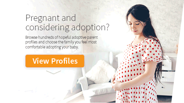
Paul & Britt
March 2005
"Miracles happen to those who believe"
We were contacted by our second MIRACLE baby’s Birth Mother from our Parent Profile. Simply AMAZING..... She said when she saw our profile "she knew we were "THE ONES". She and I "hit it off" from our first phone call. About two months later our daughter was born. We were there to attend the birth (which was AMAZING). We spent quality time with our daughter's Birth Parents and more ...
Ultrasound images are used to examine internal organs. This imaging is also called ultrasound scanning or sonography. Ultrasound imaging is achieved by sending high frequency sound waves into the body. The reflected sound waves' echoes are simply recorded and then displayed as a real time visual image.
Ultrasound images are not the same as an x-ray. Because the ultrasound images are seen in real time, they can show the movement of internal tissues and organs. This type of imaging also enables a physician to see a patient's blood flow. This ability helps diagnose a variety of conditions and makes it easier to assess possible damage caused by illnesses.
Because ultrasound images provide real time information, it can also be used to guide specific procedures such as needle biopsies in which needles are used to sample cells from organs for testing. The imaging can also help identify the source of abdominal pain or identify the cause for the enlargement of an abdominal organ.
There is not a lot of preparation needed for an ultrasound exam. The patient should wear comfortable, loose fitting clothing. There may be some other specific requirements for the ultrasound depending upon the type of imaging needed. For some ultrasound images, the doctor may request that the patient abstain from food or drink for 12 hours prior to the appointment.
Ultrasound images of the abdomen are really fast, easy, and painless. The patient will lie backside down on an examination table. The radiologist or sonographer will rub some warm gel on the patient's skin and then press the transducer firmly against the abdomen. The radiologist will move the transducer around the abdomen until the desired images are captured.
Ultrasound images are an exciting part of an expectant mother's pregnancy. The imaging is generally done at 18 to 20 weeks or at other times as may be needed. The ultrasound images show a picture of the baby on a screen. The routine exam, which takes about 20 minutes, can determine the age of the fetus, identify a possible multiple pregnancy and look for birth defects.
Ultrasound images are considered safe for all patients, including expectant mothers. When used properly, the ultrasound images provide important medical information.
More Articles: